Small-scale RUO Processing Assemblies
Static electroporation processing assemblies for research applications in small volume scales with both single-well and multi-well options.
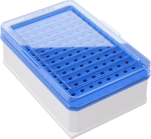
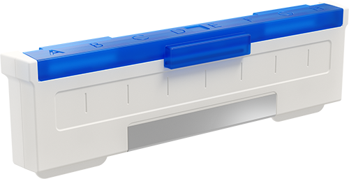
Research Use Only (RUO) Processing assemblies aid the discovery and development of applications at small scales. These static electroporation consumables are made from high-grade, inert materials to protect precious cells with additional design features for easy sample loading and recovery.
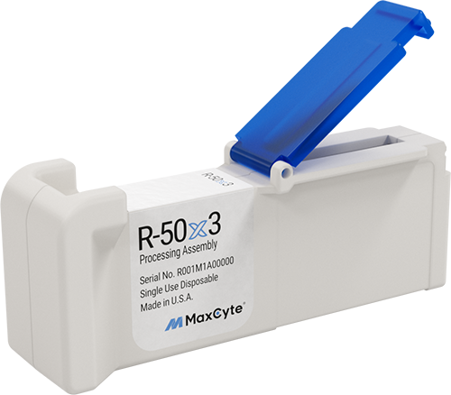
OC-100™ RUO
- Small volume applications and experimental optimization
- For quick single sample transfection
- Well designed for efficient cell loading and recovery
Configuration: Single well, 100 µL maximum volume
Cell Range: 2.5×10⁵ – 2×10⁷
Volume Range: 50 – 100 µL
Application: Research Use Only
Catalog: SOC-1
Compatible with ATx™ and STx™