CL-1.1™ RUO

Catalogue No.

CL-1.1™ RUO
  • Description
  • Graph Details

Supplier

MaxCyte

|

Catalogue No.

CL-1.1™ RUO

CL-1.1™ RUO

Mid-scale RUO Processing Assemblies
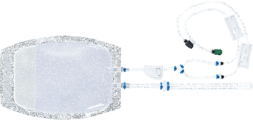
Static electroporation processing assemblies for research applications in mid-volume scales.

Research Use Only (RUO) Processing Assemblies enable the discovery and development of applications at mid-scale. These static electroporation consumables are made from high-grade, inert materials to protect precious cells with additional design features for easy sample loading and recovery.

CL-1.1™ RUO

  • Closed chamber with luer locking syringe port
  • Mid-range production scale
  • Made from high grade, inert materials to protect precious cells

Configuration: Closed chamber, 3.5 mL maximum volume
Cell Range: 5×10⁶ – 7×10⁸
Volume Range: 1 – 3.5 mL
Application: Research Use Only
Catalog: SCL-1

Compatible with ATx™ and STx™

  • Description
  • Graph Details
Description icon

Mid-scale RUO Processing Assemblies
Static electroporation processing assemblies for research applications in mid-volume scales.

Research Use Only (RUO) Processing Assemblies enable the discovery and development of applications at mid-scale. These static electroporation consumables are made from high-grade, inert materials to protect precious cells with additional design features for easy sample loading and recovery.

CL-1.1™ RUO

  • Closed chamber with luer locking syringe port
  • Mid-range production scale
  • Made from high grade, inert materials to protect precious cells

Configuration: Closed chamber, 3.5 mL maximum volume
Cell Range: 5×10⁶ – 7×10⁸
Volume Range: 1 – 3.5 mL
Application: Research Use Only
Catalog: SCL-1

Compatible with ATx™ and STx™
Graph Details icon

OC-400-v-CL-1@2x

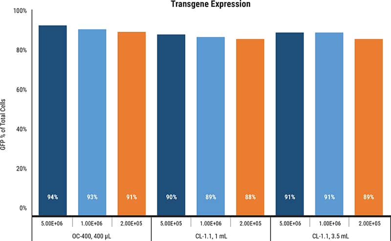
Graph Details: To make a master mix, HEK 293 cells were suspended in MaxCyte Electroporation Buffer at a density of 1×10⁸ cells/mL with pGFP at 200 µg/mL. 400 µL of the master mix was loaded into the OC-400, 1 mL was loaded into one CL-1.1 and 3.5 mL was loaded into a second CL-1.1. Transfection was performed using the MaxCyte HEK electroporation protocol for all 3 PAs. Cells were plated at 3 different densities post-transfection and were analyzed for fluorescence after 48 hours.

Related Product

SciMed (Asia) Pte Ltd | A Member of PHC Group
Privacy Overview

This website uses cookies so that we can provide you with the best user experience possible. Cookie information is stored in your browser and performs functions such as recognising you when you return to our website and helping our team to understand which sections of the website you find most interesting and useful.